
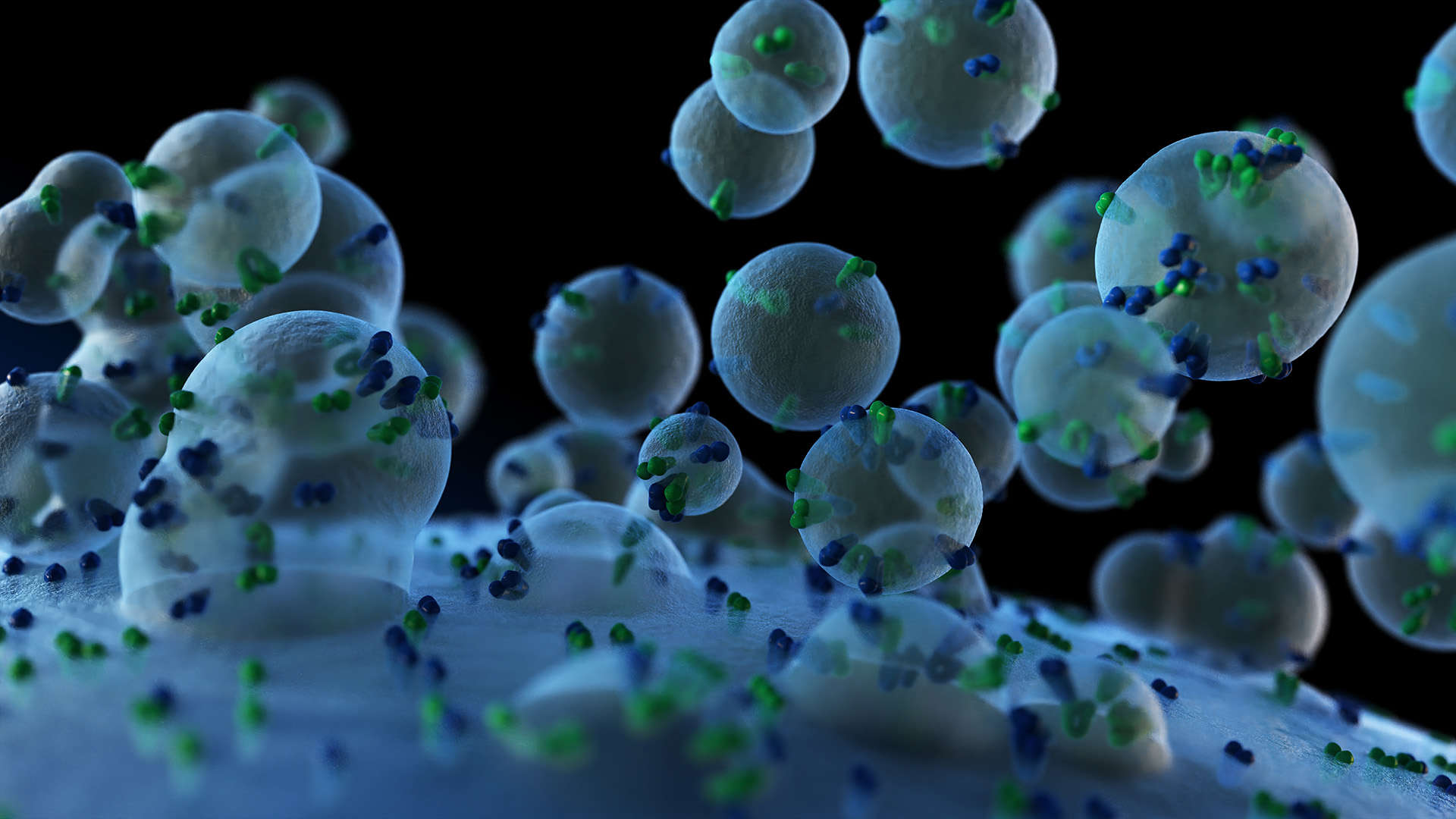
TEM-Based Exosome Identification
Online InquiryExosome formation is associated with structural modifications in the cytoplasmic membrane, a nanoscale phenomenon that is best assessed with high-resolution imaging methods. Techniques such as transmission electron microscopy (TEM) provide such resolving power and are routinely used for small intra- and extracellular structures, including exosomes. Here, we offer advanced TEM for exosome identification and analysis. Creative Proteomics aims to provide high-quality exosome identification services for customers in different fields. Our scientists help speed up your project progress based on professional knowledge and high-sensitivity equipment.
TEM-based exosome identification
As subtypes of extracellular vesicles (EVs), exosomes have a characteristic size and a unique biogenesis pattern. Exosomes are thought to be secreted by endosomal multivesicular bodies (MVB) via exocytosis and are typically ~50-100 nm in diameter. To obtain the size, concentration, composition, and other information on exosome preparation, various techniques have been developed. Among them, TEM remains the gold standard method for the visualization of EVs and is able to identify both the size and morphology of individual EVs, including exosomes.
A better understanding of the quality of your exosome samples
To ensure accurate exosome monitoring and analysis, we offer advanced TEM techniques such as cryo-electron microscopy (cryo-EM) and stain transmission electron microscopy (nsTEM). We are committed to assessing the morphology and size of exosomes from various body fluids, such as breast milk, blood plasma, urine, and saliva. Our team of experts can help researchers and professionals better identify the presence, size, and morphology of exosomes and monitor related products.
- Cryo-EM
Understanding the function of exosomes requires methods to successfully isolate and detect them without disturbing the native morphology of the vesicles. Cryo-EM has become a powerful tool for the identification of exosomes. In the process of cryo-EM, exosome preparation will be fixed and vitrified by rapid freezing in liquid ethane to preserve their original structure. By computational processing and averaging objects across multiple collected images, 3D reconstructions reaching atomic or near-atomic resolution can be generated. Cryo-EM offers unique advantages over other electron microscopes (EM), such as preserving the near-native state of exosomes, revealing the presence of lipid bilayers, and improving overall resolution.
- nsTEM
nsTEM is a technique that uses heavy metal salt solutions to embed particles into samples and enhance image contrast. Along with cryo-EM, nsTEM is a valuable tool for inspecting the purity and quality of exosome preparation, providing critical information on further processing decisions.
We can help you
- Exosome size, and size distribution
- Exosome purity, integrity, and internal morphology
- Exosome stability assessment
- Clusters/aggregates and cell debris/impurities in exosome preparation
Creative Proteomics is a leading custom service provider in the field of subcellular and single-cell research. We are committed to providing innovative solutions to power your biological research. If you have a question or want general information, please feel free to contact us. We are happy to help you.
Reference
- Noble, Jade M., et al. "Direct comparison of optical and electron microscopy methods for structural characterization of extracellular vesicles." Journal of structural biology 210.1 (2020): 107474.
* For Research Use Only. Not for use in diagnostic procedures.
