
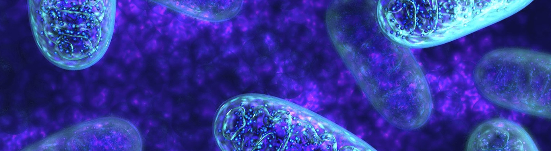
Mitochondrial Damage Detection
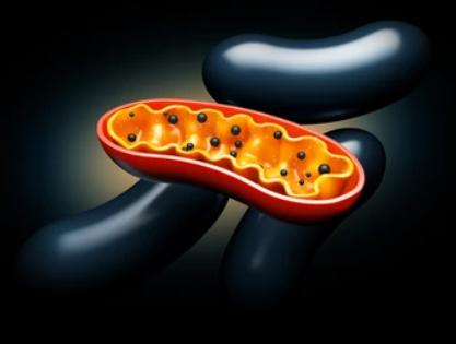
Online InquiryMitochondria are of great importance in the growth, development, metabolism, aging, disease, death and evolution of organisms. Under the action of various pathogenic factors, mitochondria are susceptible to various structural and functional damages. Mitochondrial damage is mainly manifested by increased permeability of the outer mitochondrial membrane resulting in the release of material from the membrane gap into the cytoplasm, decreased mitochondrial membrane potential, degradation of cardiolipin on the inner mitochondrial membrane or exposure of 7A6 antigen on the mitochondria. Mitochondrial damage is closely related to apoptosis. The detection of mitochondrial damage is important in the study and diagnosis of mitochondrial diseases.
Mitochondrial damage and apoptosis
The main roles of mitochondrial damage in apoptosis include the release of caspases activators, loss of electron transfer function and reduced energy production, loss of mitochondrial transmembrane potential, and the association of pro-apoptotic and anti-apoptotic functions of the BCL-2 protein family. Among them, mitochondrial release of proteins capable of activating Caspase is the most important aspect of mitochondria in the process of apoptosis. Caspase activators such as cytochrome C (Cytc), which binds with Apaf-1 and Procaspase 9 to form apoptotic bodies, thus activating Caspase 9.
Mitochondrial damage detection service
Creative Proteomics always provides you with customized services for mitochondrial damage detection services, and our one-stop research and advanced analysis platform can meet all your research needs. Our mitochondrial damage detection services include mitochondrial permeability transition pore analysis, mitochondrial membrane potential analysis, membrane phospholipid detection, and so.
- Mitochondrial permeability transition pore analysis
As a protein channel that binds the inner and outer mitochondrial membranes, mitochondrial permeability transition pore is sensitive to changes in intracellular concentrations of multiple ions, and the massive opening of mitochondrial permeability transition pores can cause disintegration of membrane potential and lead to apoptosis. Based on experienced experts and advanced platforms, such as flow cytometry, fluorescence microscopy, and laser confocal microscopy, we provides mitochondrial permeability transition pore assay services.
- Mitochondrial membrane potential analysis
The occurrence of mitochondrial damage is determined by measuring changes in intracellular mitochondrial membrane potential. Based on fluorescent detection methods and various testing platforms, we are committed to providing reliable and effective mitochondrial membrane potential assay service.
- ATP content measurement
Mitochondria are the main source of intracellular energy, and the damage to mitochondria can be determined by testing the intracellular ATP content.
- Membrane phospholipid detection
The detection of mitochondrial membrane phospholipids is important for determining the function of mitochondria, as lipid peroxidation leads to a decrease in membrane phospholipids. We mainly isolate the membrane phospholipids from mitochondria by differential centrifugation and then detect them by high performance liquid chromatography or fluorescence microscopy techniques.
- Mitochondrial morphology observation
The morphology and number of mitochondria in the cell are observed by microscopy to determine whether mitochondrial damage had occurred.
Welcome to consult for details, we will develop a detailed service agreement according to your program and needs. For more experimental technology services, please browse the rest of our website or contact us.
References
- Guo, Chunyan, et al. "Oxidative stress, mitochondrial damage and neurodegenerative diseases." Neural regeneration research 8.21 (2013): 2003.
- Abate, Marianna, et al. "Mitochondria as playmakers of apoptosis, autophagy and senescence." Seminars in cell & developmental biology. Vol. 98. Academic Press, 2020.
* For Research Use Only. Not for use in diagnostic procedures.
