
Endoplasmic Reticulum Proteomics Services
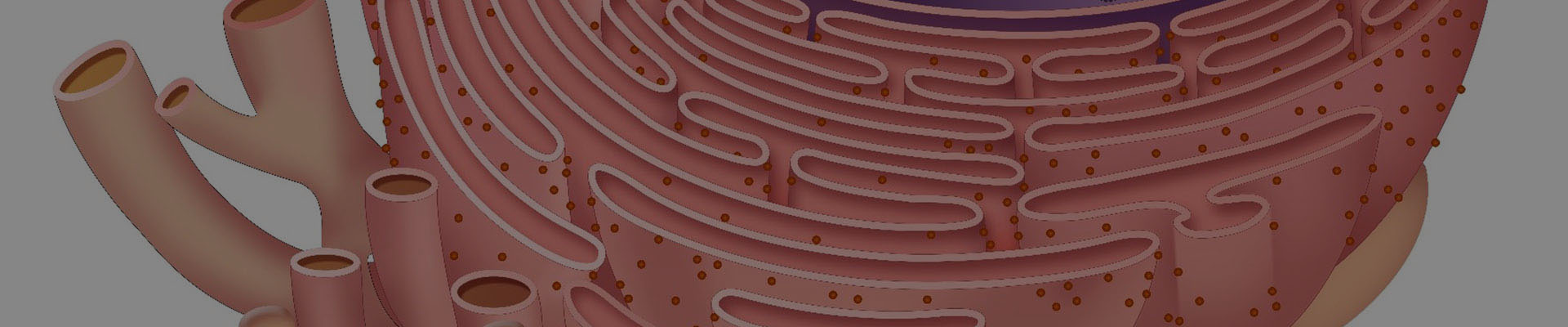
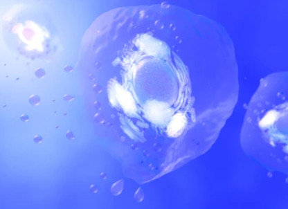
Online InquiryThe endoplasmic reticulum in eukaryotic cells is a continuous biofilm structure composed of two different morphologies: sheet and tube. The endoplasmic reticulum is involved in the synthesis of intracellular proteins and lipids and the regulation of calcium homeostasis. The lamellar endoplasmic reticulum is mainly located in the area around the nucleus of the cell, connecting inward with the outer nuclear membrane. The tubular endoplasmic reticulum is usually connected to form a network structure throughout the cytoplasm.
Creative Proteomics provides a one-stop service for endoplasmic reticulum proteomics, which aims to conduct a comprehensive analysis of endoplasmic reticulum from a holistic perspective. The main contents include: 1) Quantitative and qualitative study of endoplasmic reticulum proteins; 2) Research on the interaction between endoplasmic reticulum-related organelles; 3) Study on the maintenance of endoplasmic reticulum morphology.
We Provide the Following Services, including but not Limited to:
- Quantitative and Qualitative Study of Endoplasmic Reticulum Proteins
- Study on the Interaction Between Endoplasmic Reticulum-related Organelles
- Study on the Maintenance of Endoplasmic Reticulum Morphology
Peptide mass fingerprinting, Shotgun-MS, and top-down mass spectrometry can be used for qualitative research of proteins.
Label-based SILAC, TMT, iTRAQ quantitative technologies are available. Labeled-free SRM, PRM and SWATH technologies are available.
The endoplasmic reticulum establishes membrane contact sites with a variety of membrane cell structures through protein-protein and protein-lipid interactions, thereby performing physical activities such as material exchange, signal transduction, and membrane dynamic regulation. Defects in the interaction of the endoplasmic reticulum and membrane cells can also cause many major human diseases.
1) Study the interaction of the endoplasmic reticulum and the plasma membrane
Regulation of intracellular calcium signal.
Lipid transport and maintenance of lipid
homeostasis.
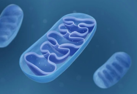
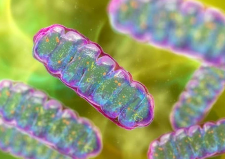
2) Study the interaction of the endoplasmic reticulum and mitochondria
Study of MAM (mitochondria-associated ER membrane) area.
Study on the relationship between the release of
calcium Ions in the endoplasmic reticulum and mitochondria.
3) Study the interaction of the endoplasmic reticulum and Golgi interaction
Study of lipid transporter-mediated lipid transport from the endoplasmic reticulum to the Golgi apparatus
4) Study the interaction of endoplasmic reticulum and endocytosis
Investigate the relationship between the contact sites of the endoplasmic reticulum and endosomes and the dynamic
changes of endosomes such as fractures and transport along microtubules
Investigate the link between the
contact sites of the endoplasmic reticulum and endosomes and the transport of cholesterol and calcium ions
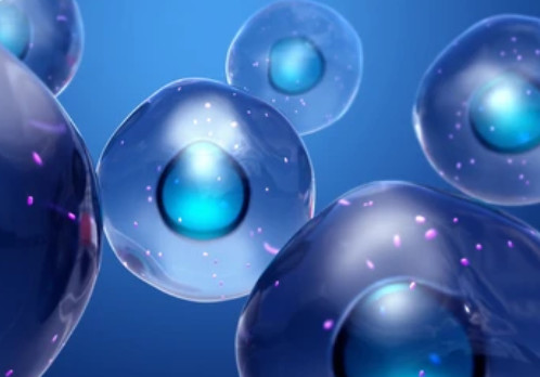
5) Study the interaction of endoplasmic reticulum and lipid droplets
Neutral lipids are synthesized on the endoplasmic reticulum membrane and gradually accumulate in the middle of the phospholipid bilayer. The new lipid droplets sprout from the endoplasmic reticulum into the cytoplasm. The lipid droplets in the cytoplasm can interact with the endoplasmic reticulum to facilitate lipid droplet growth through contact sites.
6) Study the interaction of endoplasmic reticulum and peroxisome
Study on material transport between endoplasmic reticulum and peroxisome.
The endoplasmic reticulum can be divided into two types: sheet-like and tubular. These two forms play different physiological functions.
Advantages of the Endoplasmic Reticulum Proteomics Service:
- High performance mass spectrometry system: equipped with Q Exactive Hybrid Quadrupole-Orbitrap and Agilent 6540 Q-TOF high performance mass spectrometer with multiple mass spectrometry techniques such as SRM, PRM and SWATH full scan collection, which effectively improving the identification and quantification of low abundance subcellular related proteins.
- Multi-database integration: by integrating multiple related high-quality protein databases, the accuracy and flux of subcellular protein localization can be greatly improved.
- Multi-dimensional data analysis: in addition to the comparison with the more complete related subcellular database, multiple software was used for the prediction analysis of subcellular localization based on the calculation method to further increase the identification flux.
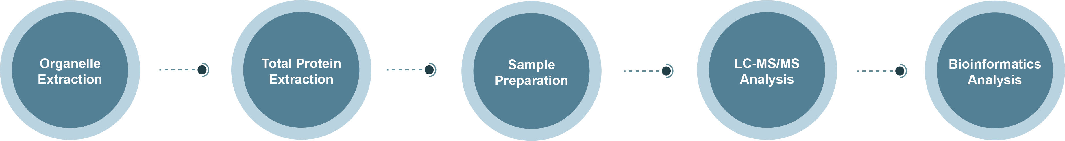
Endoplasmic Reticulum Proteomics Technology Roadmap
Data Analysis of Endoplasmic Reticulum Proteomics
| 1. Data output statistics and quality control | 2. Protein identification analysis (Density map, Scatter plot, Venn diagram) | 3. Quantitative protein analysis (Density map, Scatter plot, Venn diagram) |
| 4. Protein GO analysis | 5. Protein pathway analysis | 6. Protein COG analysis |
| 7. GO enrichment analysis of differential proteins | 8. Pathway enrichment analysis of differential proteins | 9. COG analysis of differential proteins |
References
- Itzhak D N, Sacco F, et al. SILAC-based quantitative mass spectrometry-based proteomics quantifies endoplasmic reticulum stress in whole HeLa cells. Disease Models & Mechanisms, 2019: dmm. 040741.
- Wang X, Li S, et al. Quantitative proteomics reveal proteins enriched in tubular endoplasmic reticulum of Saccharomyces cerevisiae. Elife, 2017, 6: e23816.
Related Services
* For Research Use Only. Not for use in diagnostic procedures.
