
Mitochondrial Autophagy Research
Online InquiryMitochondrial autophagy (mitophagy) is a biological phenomenon that occurs in cells in response to malnutrition or external stimulation. Mitophagy can selectively remove or degrade damaged mitochondria to maintain intracellular homeostasis, which is critical for the functional integrity of the entire mitochondrial network and cell survival. Abnormal mitochondrial autophagy exacerbates mitochondrial dysfunction and is closely associated with the development of many diseases. Mitochondrial autophagy is receiving more and more attention. To help our clients study mitochondrial autophagy more efficiently, we offer a one-stop mitochondrial autophagy research service.
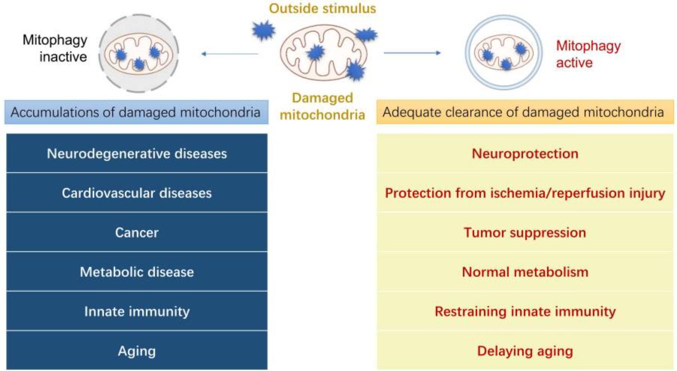 Fig.1 Physiological functions of mitophagy in human diseases. (Lu, Yingying, et al, 2023)
Overview of mitochondrial autophagy
Mitochondria are involved in ATP production, calcium homeostasis, metabolic synthesis, oxidative stress response, and apoptosis. Dysfunctional or impaired mitochondrial function may lead to serious consequences, even to cell death. Mitophagy is a defense mechanism that selectively removes damaged and dysfunctional mitochondria in cells. Increasing mitophagy under different stress conditions can maintain cellular ATP levels, reduce ROS-induced oxidative damage, and selectively remove damaged mitochondria from the cell, thereby playing an essential role in mitochondrial quality control.
Mitochondrial autophagy classification
- Basic mitochondrial autophagy: cells continuously clean up senescent and damaged mitochondria, ensuring that they can be recycled. Organs with high levels of mitophagy include: heart, liver, kidney, skeletal muscle and nervous system.
- Stress-induced mitochondrial autophagy: Extracellular stress signals affect the physiological function of mitochondria and cause depletion of mitochondrial membrane potential, leading to acute mitochondrial clearance.
- Programmed mitochondrial autophagy: Programmed mitophagy is activated during the development of different cells, such as retinal ganglion cell development, chemical reprogramming process of somatic cells to pluripotent stem cells, maturation of cardiomyocytes, erythroid differentiation, and mitochondrial clearance of spermatozoa origin after fertilization.
Mitochondrial autophagy research service
Creative Proteomics is based on advanced analytical platforms and tools for mitochondrial autophagy research that help our customers accurately observe and track the dynamics of mitochondrial autophagy in real time. Since mitophagy is a complex and dynamic process, detection methods are constantly updated. We offer the following services, but are not limited to:
- Mitochondrial function detection
-Mitochondrial membrane potential analysis
-Mitochondrial calcium ion detection
-Mitochondrial ROS analysis
-Mitochondrial ATP content determination
- Mitochondrial morphology observation
Transmission electron microscopy (TEM) is used to observe damaged mitochondria and autophagosomes with mitochondria-like organelles.
- Mitochondrial autophagy marker assay
Mitochondrial autophagy markers such as PINK1, Parkin, p62, Beclin1, LC3B.
- Induction and inhibition of mitochondrial autophagy
In general, mitochondrial autophagy is induced or inhibited by human intervention through drug treatment, physical damage, starvation, knockdown or overexpression of mitochondrial autophagy genes, etc. Cyclosporin A is a drug that inhibits altered mitochondrial permeability and acts as a protective agent after induction of mitochondrial autophagy, significantly reducing the total amount of mitochondrial autophagosomes.
- Pathways of mitochondrial autophagy
-PINK1/Parkin-mediated mitophagy
-BNIP3 and BNIP3L/Nix-mediated mitophagy
-FUNDC1-mediated mitophagy
-Lipid-mediated mitophagy
Our mitophagy research platform
- Electron microscopy (EM)
- Fluorescence microscopy (FM)
- Immunofluorescence microscopy (IF)
- Immunoblotting (IB)
- Flow cytometry (FC)
Creative Proteomics excels at handling challenging projects, and our scientists have a wide range of expertise and experience in mitochondrial autophagy research. Would you like more information about our services? Please contact us.
References
- Lu, Yingying, et al. "Cellular mitophagy: Mechanism, roles in diseases and small molecule pharmacological regulation." Theranostics 13.2 (2023): 736.
- Li, Anqi, et al. "Mitochondrial autophagy: Molecular mechanisms and implications for cardiovascular disease." Cell Death & Disease 13.5 (2022): 444.
* For Research Use Only. Not for use in diagnostic procedures.
