
Mitochondrial Phenotype Analysis using Electron Microscopy
Online InquiryTransmission electron microscopy (TEM) has long been an important technique for the high-resolution and visualization of subcellular structures within cells and tissues. This powerful tool is able to afford high-resolution images by transmitting electrons through an ultrathin portion of the sample and magnifying the resulting image using a series of lenses. As a leading service provider in mitochondrial morphology analysis, Creative Proteomics is proud to offer TEM to study ultrastructural changes of mitochondria in a wide range of cells and tissues. Our services can help researchers and professionals to observe the ultrastructure of mitochondria under normal and disease conditions, visualize fission/fusion and mitophagy, etc.
Overview of mitochondrial ultrastructure
Mitochondria are primarily responsible for the production of ATP, which is required for all energy-dependent cellular processes. As a major energy organelle, mitochondria produce ATP through oxidative phosphorylation of the inner mitochondrial membrane, and thus their function is closely related to mitochondrial ultrastructure. The outer and inner mitochondrial membranes enclose and define the intermembrane space and matrix compartments. The invaginated intermembrane forms cristae, where the respiratory chain complex is located. Structural changes that accumulate in mitochondria with age contribute to the regulation of reduction-oxidation homeostasis and can lead to a decline in mitochondrial function. Disturbances in the ultrastructure of the mitochondrial cristae may lead to defects in ATP synthesis. In addition to producing less ATP, damaged and poorly functioning mitochondria can induce the formation of cytotoxic reactive oxygen species (ROS).
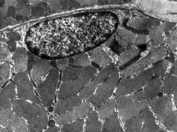
Mitochondrial phenotype analysis using TEM
Mitochondria play an essential role in multiple metabolic pathways (such as apoptosis and cell death pathways) and are responsible for the generation of cellular energy. TEM is a straightforward and powerful method to quantify and characterize mitochondria and their ultrastructures. Creative Proteomics has developed an advanced TEM platform. Based on this technique, we are able to help researchers and professionals to study the ultrastructure of mitochondria in a wide variety of healthy and diseased cells and tissues, providing important insights into cellular processes and disease development. Our TEM-based mitochondrial morphology analysis service consists of four main parts, including experimental design, TEM sample preparation, TEM analysis, and morphological and statistical analyses from TEM images.
Specifically, our services can help our clients with the following lists:
- Mitochondrial morphology analysis
- Mitochondrial length and width measurement
- Cristae morphology analysis
- Mitochondria-other organelle contact analysis
As a major energy organelle, mitochondria play a role in many complex diseases, such as Alzheimer's disease, diabetes, and cardiomyopathy. Understanding how these diseases affect mitochondrial structure by using enhanced TEM can lead to a better understanding of their pathophysiology. Whether you need sophisticated analytical services to analyze mitochondrial morphology or a broader interpretation of analytical data, our experienced expert team is committed to meeting your needs. If you have any further questions about our platform or services, please contact us, and our experienced electron microscopy experts will help you.
Reference
- Lam, Jacob, et al. "A universal approach to analyzing transmission electron microscopy with ImageJ." Cells 10.9 (2021): 2177.
* For Research Use Only. Not for use in diagnostic procedures.
